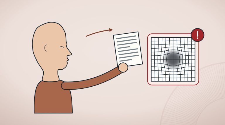
For a few months, sometimes a few years, everything went as hoped: after the operation vision was sharp again and reading was easier. Then, slowly, the veil that people thought they had left behind in the operating theatre came back. At that point, many people think the cataract has returned.
In fact, the cloudy lens was removed during the operation. What becomes hazy is a very thin envelope left in the eye on purpose to hold the artificial lens: the posterior capsule. Ophthalmologists speak of posterior capsule opacification; in everyday language it is secondary cataract, the most frequent complication of cataract surgery1,2.
In brief
- Secondary cataract is not a return of the cataract: it is the clouding of the capsule that holds the artificial lens, caused by cells left behind after the operation1.
- It is common: in a British study of more than 20,000 eyes, at five years laser capsulotomy had been needed in a share of roughly 6 to 19%, depending on the lens model3.
- It is treated with a YAG laser, which opens a gap in the cloudy capsule1,2; the procedure is quick and non-invasive2.
- After the laser, eye pressure may rise or macular oedema may appear; retinal detachment is rare6.
- Lenses with a square posterior edge are associated with fewer capsulotomies over the years9.
In this article
What secondary cataract is
In the operation, the cloudy content of the lens is removed, while the transparent bag that surrounded it, the capsule, stays in place and becomes the home of the artificial lens (we describe this in our article on cataract surgery). Some cells from the lens, however, can remain on the capsule.
A 2009 review describes what happens next: these residual cells can multiply, migrate towards the centre of the posterior capsule and transform, depositing collagen and forming new fibres1. The result is a cloudy screen right behind the artificial lens, which once again blurs images as the cataract did. Surgical techniques and lens materials have reduced the problem, but have not eliminated it1.
How common secondary cataract is
Estimates vary a great deal from one study to another, partly because different definitions are used. A French review from 2014 speaks of an incidence of around 30% and observes that the opacification tends to be regarded almost as a natural stage in the history of the operation2.
The most solid data come from clinical practice. A British study analysed the electronic records of 20,763 eyes of people aged 65 or over, operated on between 2010 and 2013 in seven public health service centres with monofocal acrylic lenses. Three years after the operation, laser capsulotomy had been needed in between 2.4 and 12.6% of eyes, and at five years in between 5.8 and 19.3%, depending on the lens model implanted3. The study, which is observational, shows differences between the lenses but cannot rule out that they depend in part on the patients or the centres.
In France, a study based on the representative sample of the national reimbursement database estimated about 312,000 capsulotomies in 2017 alone, in people with an average age of 75. Among those who had been treated, one person in three had been treated within two years of the cataract operation and about one in ten within the first year4. It is therefore a common event, which can appear early but also a long time afterwards.
How it is treated: Nd:YAG laser capsulotomy
According to the 2009 review, the only effective treatment for secondary cataract is Nd:YAG laser capsulotomy1. The ophthalmologist aims the laser at the cloudy capsule and opens a small gap in its centre, so that light can reach the retina unobstructed again. There is no need to return to the operating theatre or to open the eye: the French review calls it a quick and non-invasive procedure, while warning that opening the capsule can be associated with a number of complications2. For this reason, the authors conclude, the preferable route remains preventing the opacification2. The recommendations of the European Society of Cataract and Refractive Surgeons (ESCRS) also list it among the possible events after the operation5.
The risks of the laser, in numbers
The study
YAG 2: what happens in the year after capsulotomy
A French observational study followed for 12 months, through the representative sample of the national reimbursement database, 6,210 adults treated with YAG capsulotomy between 2014 and 2017, for a total of 7,958 procedures; people with eye diseases in the previous year were excluded. At three months, 8.6% had had at least one of the adverse events studied, and at one year 13.3%. The most frequent were a rise in eye pressure and macular oedema, a build-up of fluid in the macula, the central area of the retina: each around 5% in the first three months. Retinal detachment stayed below 0.5% throughout the period. The risk was higher in people with diabetes and in those who had been treated within a year of the operation6. The main limitation: an administrative database records coded diagnoses and does not say how much each event affected vision.
The most frequently cited fear is retinal detachment, the lifting of the light-sensitive layer from the inner wall of the eye. Here the studies do not all point in the same direction. A 2022 meta-analysis of 11 studies and 65,117 eyes operated on for cataracts found a higher risk of detachment after capsulotomy: a relative risk of 1.57, that is, about 57% more than in those who had not had it7. The association, however, was clear mainly in Asian studies and after the old extracapsular technique, whereas it did not emerge in studies on phacoemulsification, the ultrasound technique7.
A study of 17,688 eyes operated on with phacoemulsification in a Finnish hospital, followed for a little over four years on average, counted 83 retinal detachments, equal to 0.11% a year. Once age, sex and the power of the implanted lens had been taken into account, capsulotomy was not associated with a higher risk; what did count were male sex, younger age and a lower lens power8.
Can the risk be reduced? The role of the lens
No lens removes the risk, but some characteristics seem to reduce it. The most studied is the shape of the edge of the optical part. A 2021 Cochrane review pooled 10 randomised trials (1,065 people, 1,834 eyes), in eight of which one eye received a square-edged lens and the other a round-edged one. At one year capsulotomies were few in both groups (1 in 371 against 4 in 371) and the result was imprecise; as the years passed the difference became clearer: at three years, 170 fewer capsulotomies for every 1,000 square-edged lenses9.
The material matters too. A 2017 meta-analysis of 11 studies and 889 eyes compared hydrophobic acrylic lenses, which repel water, with hydrophilic acrylic ones: with the former, the likelihood of needing the laser was lower, while the final vision was similar10. The choice of lens is up to the surgeon; we describe the types in our article on artificial lenses.
When to talk to an ophthalmologist
- If, months or years after cataract surgery, your vision slowly becomes veiled again: this is a good reason for an examination, because secondary cataract can be treated and has to be distinguished from other causes1,5.
- If in the weeks after a capsulotomy your vision worsens again: among the most frequent events are a rise in eye pressure and macular oedema6.
- Immediately, the same day: a drop in vision after an initial improvement, the sudden appearance of black dots, flashes of light, or increasing pain or redness in the operated eye are the signs for which the ESCRS advises asking for help without waiting5. Call 112 or go to an emergency department (A&E); you will find the symptoms of retinal detachment and the other warning signs on our page about eye emergencies.
Frequently asked questions
Can a cataract come back after surgery?
Strictly speaking, no, because the cloudy lens is removed. What can happen is clouding of the capsule that holds the artificial lens, so-called secondary cataract, which is the most frequent complication of the operation1,2.
How long after surgery does secondary cataract appear?
It can appear after months or after years. In the French study, among the people treated with the laser, about one in ten had been treated within the first year and one in three within two years4; in the British study the cases kept increasing between the third and the fifth year3.
Is the YAG laser dangerous?
It is a quick, non-invasive procedure2, but not free of undesirable effects: in a large French study, in the following three months, about 5% of people had a rise in eye pressure and a similar share had macular oedema, while retinal detachment stayed below 0.5%6.
Can secondary cataract be avoided?
Not always. Lenses with a square posterior edge and those made of hydrophobic acrylic are associated with fewer capsulotomies over time9,10, but the risk does not disappear. The choice of lens is made with the surgeon.
When vision becomes hazy again after a cataract has been operated on, the explanation is often not a failed operation or a disease coming back, but a capsule that has become veiled, the most frequent complication of this surgery1. It is a known problem, treated with a laser; knowing its risks, small but real, helps to explain why, after a capsulotomy, the ophthalmologist may want to see the eye again.
Sources
- Awasthi N, Guo S, Wagner BJ. Posterior capsular opacification: a problem reduced but not yet eradicated. Arch Ophthalmol. 2009;127(4):555-62. doi:10.1001/archophthalmol.2009.3 · PubMed 19365040
- Milazzo S, Grenot M, Benzerroug M. [Posterior capsule opacification]. J Fr Ophtalmol. 2014;37(10):825-30. doi:10.1016/j.jfo.2014.09.003 · PubMed 25455552
- Ursell PG, Dhariwal M, O’Boyle D, Khan J, Venerus A. 5 year incidence of YAG capsulotomy and PCO after cataract surgery with single-piece monofocal intraocular lenses: a real-world evidence study of 20,763 eyes. Eye (Lond). 2020;34(5):960-968. doi:10.1038/s41433-019-0630-9 · PubMed 31616057 · PMC
- Brézin AP, Labbe A, Schweitzer C, Lignereux F, Rozot P, Goguillot M, et al. Incidence of Nd:YAG laser capsulotomy following cataract surgery: a population-based nation-wide study – FreYAG1 study. BMC Ophthalmol. 2023;23(1):417. doi:10.1186/s12886-023-03134-6 · PubMed 37845645 · PMC
- European Society of Cataract and Refractive Surgeons (ESCRS). ESCRS Guideline for Cataract Surgery. 2024. escrs.org
- Dot C, Schweitzer C, Labbé A, Lignereux F, Rozot P, Goguillot M, et al. Incidence of Retinal Detachment, Macular Edema, and Ocular Hypertension after Neodymium:Yttrium-Aluminum-Garnet Capsulotomy: A Population-Based Nationwide Study-The French YAG 2 Study. Ophthalmology. 2023;130(5):478-487. doi:10.1016/j.ophtha.2022.12.022 · PubMed 36581227
- Liu H, Liu X, Chen Y, Wang D, Li Y, Chen H, et al. Effect of Nd:YAG laser capsulotomy on the risk for retinal detachment after cataract surgery: systematic review and meta-analysis. J Cataract Refract Surg. 2022;48(2):238-244. doi:10.1097/j.jcrs.0000000000000755 · PubMed 34538778
- Elbaz U, Hakkala L, Hecht I, Achiron A, Gershoni A, Tuuminen R. Nd:YAG capsulotomy is not a risk factor for retinal detachment after phacoemulsification cataract surgery. Acta Ophthalmol. 2021;99(7):e1018-e1026. doi:10.1111/aos.14757 · PubMed 33423371
- Maedel S, Evans JR, Harrer-Seely A, Findl O. Intraocular lens optic edge design for the prevention of posterior capsule opacification after cataract surgery. Cochrane Database Syst Rev. 2021;8(8):CD012516. doi:10.1002/14651858.CD012516.pub2 · PubMed 34398965 · PMC
- Zhao Y, Yang K, Li J, Huang Y, Zhu S. Comparison of hydrophobic and hydrophilic intraocular lens in preventing posterior capsule opacification after cataract surgery: An updated meta-analysis. Medicine (Baltimore). 2017;96(44):e8301. doi:10.1097/MD.0000000000008301 · PubMed 29095259 · PMC
Study titles are given in their original language of publication. The superscript numbers in the text refer to these sources; citation details come from PubMed (National Library of Medicine).