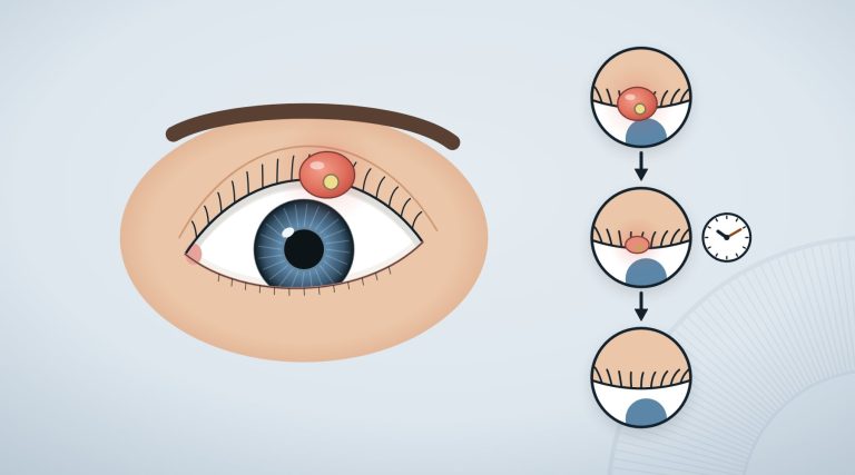
It shows up in photographs before it does in the mirror: one eye looks smaller than the other, the gaze more tired. Sometimes the upper eyelid droops so far that it covers part of the pupil, and to see well you raise your chin or lift your eyebrows without realising.
A drooping eyelid is called in medicine eyelid ptosis, or blepharoptosis. It is one of the most common eyelid problems in ophthalmology clinics2, and its causes range from a tendon that loosens with the years to diseases of the nerves and muscles. For this reason, before ‘how it is corrected’, comes ‘why it happened’.
In brief
- A drooping eyelid, or ptosis, can be present from birth or appear later; the acquired form is the most frequent2.
- In adults the causes range from age-related changes to rigid contact lenses, eye surgery and diseases such as myasthenia3,4,5,6.
- An eyelid that drops suddenly with double vision, a dilated pupil or pain can be a neurological emergency: call 1127.
- In children congenital ptosis needs to be followed, because it can lead to lazy eye (amblyopia)1,8.
- Surgery is the main treatment, and two techniques compared give a similar final height; an eye drop studied recently lifts the eyelid by less than a millimetre3,9,10.
In this article
What a drooping eyelid (ptosis) is
It is called ptosis when the edge of the upper eyelid sits lower than normal. It can be mild (1–2 millimetres), moderate (3–4 millimetres) or severe, when it exceeds 4 millimetres and comes to cover the whole pupil. It can affect one eye or both1.
Besides the tired look, ptosis can cause blurred vision and watering. People with a marked one tend to hold the head back with the chin raised, to lift the eyelid with a finger or to raise the eyebrows; the continuous effort of the forehead muscles can cause headache and eye strain1. The upper part of the visual field is often lost, with an effect on quality of life2,3.
Two muscles mainly lift the eyelid: the levator muscle and a small muscle called Müller’s muscle3. The causes of ptosis are divided according to what is not working: the muscle (myogenic causes), the nerve (neurogenic), the tendon that connects the levator to the eyelid (aponeurotic), a weight or mechanical obstacle, or an injury1.
The causes of a drooping eyelid in adults
Age. Among the common causes are the changes linked to ageing3.
Contact lenses. A 2015 systematic review with meta-analysis pooled 5 studies on over 7,400 people: in people who wore rigid contact lenses the odds of having ptosis were about 17 times higher than in those who did not. It is a relative estimate and a very imprecise one (the confidence interval ranged from about 4 to over 80); for soft lenses there was only one study, on 90 people, which also showed a higher risk4. The authors suggest always asking whether a person with acquired ptosis wears or has worn contact lenses4. The differences between types of lenses are explained in the article on contact lenses.
Eye surgery. According to a meta-analysis of 16 studies on 2,856 eyes, ptosis appeared after eye surgery in about 11 cases in 100, more often in women; the type of operation mattered5.
Diseases of the nerves and muscles. Ptosis can be part of general diseases, such as myasthenia gravis or Horner syndrome3. Myasthenia is an autoimmune disease in which the signal between nerve and muscle passes poorly: its typical sign is fluctuation, that is, an eyelid that comes and goes, gets worse with tiredness and is often accompanied by double vision6. In Horner syndrome, described in the article on heterochromia, the slightly drooping eyelid comes with a smaller pupil on the same side. Double vision and its causes have a separate article.
When a drooping eyelid is an emergency
There are two situations in which an eyelid that suddenly drops needs to be assessed at once, because it can be the sign of a problem of the blood vessels of the brain or neck. An overview of neuro-ophthalmic emergencies indicates urgent MRI7:
- when the eyelid drops together with painful double vision and a dilated pupil, to rule out an aneurysm, a dilation of an artery of the brain;
- when a Horner syndrome, with a slightly drooping eyelid and a smaller pupil, comes with pain, to rule out a dissection of the internal carotid artery, a tear in the wall of the artery of the neck.
In these cases you do not wait for the scheduled appointment: you call 112 or go to an emergency department (A&E).
Ptosis in children
Congenital ptosis appears within the first year of life, on its own or as part of other eye or general diseases8. The critical point is vision: if the eyelid covers the pupil, or if it comes with an astigmatism that differs between the two eyes, the child can develop amblyopia, lazy eye, with reduced vision that can become permanent1,8.
Not all children with congenital ptosis need surgery, but those with amblyopia can benefit from early correction8. Surgery in children is not simple, and more than one operation is sometimes needed8. How lazy eye is recognised and treated is described in the article on amblyopia in children.
How it is corrected: surgery and eye drops
The choice depends on age, on the cause, on how far the eyelid drops and on how much strength the levator muscle still has. The options range from watchful waiting to drugs, up to surgery1, which remains the main approach3.
For mild ptosis, operations are used that shorten Müller’s muscle from inside the eyelid; when the levator still works, it is shortened or repositioned; when it works little, the eyelid is connected to the forehead muscle, which lifts it in its place1. Among the risks, uncommon, are infection, bleeding, over- or under-correction and, rarely, a drop in vision; in the early period it can be difficult to close the eye completely, and the two eyelids do not always turn out perfectly symmetrical1.
The study
Two techniques compared
A 2023 systematic review with meta-analysis pooled 7 comparative studies, with 1,038 operated eyelids, that compared shortening of Müller’s muscle from inside the eyelid with repositioning of the levator tendon from outside. The final height of the eyelid was similar. With the second technique, however, over-corrections and further operations were more frequent, while the first took on average about 11 minutes less. The main limitation: there are only 7 studies9.
For some years an eye drop has also been studied, oxymetazoline, a drug that stimulates adrenergic receptors. In a meta-analysis of 4 randomised trials on 448 people with acquired ptosis, the eyelid lifted on average by 0.65 millimetres with the eye drop against 0.26 with the placebo, and serious adverse events were rare and similar in the two groups (1.2% and 1.6%)10. It is a measurable but small effect, under a millimetre.
When to talk to an ophthalmologist
- An eyelid that droops little by little, on one or both sides, especially if it covers the pupil or if you raise your chin to see: talk to the ophthalmologist1.
- An eyelid that drops more in the evening or with tiredness, perhaps with double vision that comes and goes: it can suggest myasthenia, to be assessed soon6.
- A newborn or child with an eyelid that partly covers the pupil: have it seen by the ophthalmologist, because it can lead to lazy eye1,8.
- Immediately, by calling 112: an eyelid that drops suddenly with double vision, a dilated pupil or severe headache, or with a smaller pupil and pain in the neck, face or head7. You will find the other warning signs on our page about eye emergencies.
Frequently asked questions
Why does only one of my eyelids droop?
The possible causes are many: age, contact lenses, eye surgery, diseases of the nerves or muscles such as myasthenia3,4,5,6. If the eyelid drops suddenly, with double vision, a pupil different from the other or pain, it is an emergency7.
Can contact lenses make the eyelid droop?
According to a meta-analysis, people who wear rigid contact lenses have many times higher odds of ptosis than those who do not, although the estimates are imprecise; a higher risk was also observed with soft lenses, in a single study4.
Is surgery for ptosis risky?
Complications are uncommon: infection, bleeding, over- or under-correction and, rarely, a drop in vision; and the two eyelids do not always turn out perfectly symmetrical1. With some techniques a further operation may be needed more often than with others9.
Are there eye drops for a drooping eyelid?
An oxymetazoline eye drop has been studied in people with acquired ptosis: in the meta-analysis of randomised trials it lifted the eyelid on average by 0.65 millimetres, against 0.26 with the placebo10. Whether it can help in a specific case, and whether it is available where you live, is for the ophthalmologist to assess.
A drooping eyelid is often a sign of age, which can be corrected when it bothers you1,3. First, however, it matters to understand where it comes from: ptosis that comes and goes, or that arrives suddenly with other problems, needs to be assessed soon6,7.
Sources
- Finsterer J. Ptosis: causes, presentation, and management. Aesthetic Plast Surg. 2003;27(3):193-204. doi:10.1007/s00266-003-0127-5 · PubMed 12925861
- Bacharach J, Lee WW, Harrison AR, Freddo TF. A review of acquired blepharoptosis: prevalence, diagnosis, and current treatment options. Eye (Lond). 2021;35(9):2468-2481. doi:10.1038/s41433-021-01547-5 · PubMed 33927356 · PMC
- Chung CW, Gandhi PD. Ptosis Evaluation for Otolaryngologists. Otolaryngol Clin North Am. 2025;58(5):859-872. doi:10.1016/j.otc.2025.03.018 · PubMed 40374478
- Hwang K, Kim JH. The Risk of Blepharoptosis in Contact Lens Wearers. J Craniofac Surg. 2015;26(5):e373-4. doi:10.1097/SCS.0000000000001876 · PubMed 26102541
- Wang Y, Lou L, Liu Z, Ye J. Incidence and risk of ptosis following ocular surgery: a systematic review and meta-analysis. Graefes Arch Clin Exp Ophthalmol. 2019;257(2):397-404. doi:10.1007/s00417-018-4130-6 · PubMed 30203103
- Keene KR, Kan HE, van der Meeren S, Verbist BM, Tannemaat MR, Beenakker JM, et al. Clinical and imaging clues to the diagnosis and follow-up of ptosis and ophthalmoparesis. J Cachexia Sarcopenia Muscle. 2022;13(6):2820-2834. doi:10.1002/jcsm.13089 · PubMed 36172973 · PMC
- Heran F, Schaison-Cusin M, Williams M, Piekarski JD. [Imaging of neuro-ophthalmological emergencies]. J Neuroradiol. 2004;31(4):291-300. doi:10.1016/s0150-9861(04)97008-3 · PubMed 15545941
- SooHoo JR, Davies BW, Allard FD, Durairaj VD. Congenital ptosis. Surv Ophthalmol. 2014;59(5):483-92. doi:10.1016/j.survophthal.2014.01.005 · PubMed 24657037
- Karam M, Alsaif A, Abul A, Alkhabbaz A, Alotaibi A, Shareef E, et al. Muller’s muscle conjunctival resection versus external levator advancement for ptosis repair: systematic review and meta-analysis. Int Ophthalmol. 2023;43(7):2563-2573. doi:10.1007/s10792-023-02633-1 · PubMed 36692699
- Bawazir RO, Qedair JT, Bukhari ZM, Badghaish OS, Hubayni RA, Alsudais AS, et al. Efficacy of Oxymetazoline 0.1% in Acquired Blepharoptosis: A Systematic Review and Meta-Analysis of Randomized Clinical Trials. Clin Ophthalmol. 2025;19:2955-2964. doi:10.2147/OPTH.S529633 · PubMed 40881581 · PMC
Study titles are given in their original language of publication. The superscript numbers in the text refer to these sources; citation details come from PubMed (National Library of Medicine).