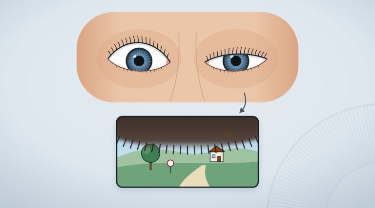
It usually happens without warning. You are reading and you can no longer see out of one eye, or you see as if through a dark veil. No pain, no redness. Sometimes it lasts a few minutes and then everything returns to normal, and the temptation is to forget about it.
It is precisely in these cases that specialists ask people to behave as they would faced with a stroke. A sudden, painless loss of vision in one eye can be the sign that an artery of the retina has closed, an event that the most recent reviews regard as the equivalent of a stroke of the brain1. The same applies to blurred vision in one eye that appears suddenly and has no obvious explanation.
In brief
- A sudden, painless loss of vision in one eye can be due to occlusion of an artery of the retina, regarded as the equivalent of a stroke1.
- It is an emergency for the brain too: in a meta-analysis, 30 people in 100 with central retinal artery occlusion had a recent cerebral ischaemia visible on MRI3.
- Even when vision returns after a few minutes (amaurosis fugax) an urgent assessment is needed, because the risk of stroke is highest in the first days4,6.
- Over 50, giant cell arteritis should be considered, an inflammation of the arteries that leads to loss of vision in about 18 people in 100 among those affected7.
- The advice is the same as for stroke: call 1121,10.
In this article
The eye stroke: central retinal artery occlusion
The retina receives blood mainly from one small vessel, the central retinal artery, which enters the eye together with the optic nerve. When it closes, vision drops suddenly, in one eye only and usually without pain1. For this reason the American Heart Association, in a 2021 scientific statement, defines it as a medical emergency and asks that emergency systems recognise it and direct it at once to urgent care2.
The diagnosis is made by examining the back of the eye, with the help of photographs of the retina and OCT, a test that shows its layers1. What can be done afterwards is still a subject of research. There is no therapy accepted by everyone, and traditional treatments, such as massaging the eye or drawing off a little fluid from the eye (paracentesis), have shown no benefit and should not be used1.
Hopes centre on thrombolysis, the clot-dissolving medicines used in stroke of the brain: recent reviews suggest a possible effect if given intravenously within 4.5 hours of the loss of vision, or into an artery within 6 hours, and randomised trials are under way to get a clear answer1,2.
The study
Thrombolysis: what the data from 783 patients say
A meta-analysis published in 2025 collected individual patient data from 35 studies: 783 people with severe vision loss due to central retinal artery occlusion, treated within 24 hours. The main outcome was recovering from the severe vision loss. This was achieved by 28.8% of those who had received intravenous thrombolysis within 4.5 hours, against 11.1% of those who had only traditional therapies; with intra-arterial thrombolysis within 6 hours, 27.2% against 12.0%. Each hour gained was associated with a greater improvement. The authors warn that the results need to be confirmed by randomised trials that are large enough: by their calculations 95 people per group would be needed5.
For the reader the point is not the therapy, which is for the doctors, but time: windows of a few hours are easily lost by waiting for vision to return on its own.
Why you go to a stroke centre
A closed artery of the retina is not only a problem of the eye. A 2020 meta-analysis pooled the studies in which people with a retinal arterial occlusion had had a brain MRI within 7 days: a recent cerebral ischaemia was present in 30% of those with a central artery occlusion and in 25% of those with a branch occlusion3. In many cases it caused no neurological symptoms: silent ischaemias involved 22% of people with central artery occlusion3.
A 2018 review stresses that the risk of stroke is highest in the first days after the loss of vision. For this reason guidelines recommend an urgent MRI of the brain and an examination of the vessels, and ask eye specialists to send these people at once to a stroke centre, without wasting time on other tests4. Risk factors for vascular disease must also be sought and treated quickly2. And for someone with the symptom, the most useful thing is to call the emergency services, so as to be taken directly to a hospital with a stroke centre1.
When vision returns: amaurosis fugax
If vision darkens in one eye and then returns within minutes, we speak of transient monocular vision loss, or amaurosis fugax. It has many causes, but most often it is due to a temporary lack of blood to the retina: it can herald permanent loss of vision or a stroke, and for this reason must be assessed urgently6. In the 2020 meta-analysis, almost 12 people in 100 with this problem already had a recent cerebral ischaemia on MRI3.
The UK National Health Service (NHS) too lists, among the symptoms of stroke, blurred or lost vision in one or both eyes, and recalls that symptoms can pass quickly: to those who have had them in the last 24 hours it nevertheless advises asking for help at once10. In younger people amaurosis fugax is often benign, but the assessment must in any case be adapted to each case6. We discuss the other causes of hazy vision, more or less urgent, in Sudden blurred vision.
Over 50: giant cell arteritis
In older people there is a cause not to be forgotten: giant cell arteritis, an inflammation of the large arteries, the most common vasculitis after the age of 507. A 2026 review reports that it leads to a loss of vision in about 18% of cases, and that this loss can be avoided if the disease is recognised, investigated and treated early7. Treatment with corticosteroids is effective, although almost half of people have a relapse7.
The typical signs are headache and visual disturbances, up to loss of vision; in blood tests the markers of inflammation are high in over 9 people in 108. The diagnosis is confirmed with a biopsy of the temporal artery, while ultrasound and MRI of the vessels help to support it8. For this reason, faced with a sudden drop in vision in an older person, doctors look for this disease too6.
The other causes to recognise at once
A 2026 review that proposes to doctors a pathway in five steps recalls that acute glaucoma attack, retinal detachment and optic neuritis must also be recognised immediately, as well as retinal artery occlusion and giant cell arteritis9. The first two questions of that pathway are simple: which eye is affected, and whether there is pain9. We have described the symptoms of retinal detachment and the difference between open-angle and angle-closure glaucoma in dedicated articles.
When to talk to an ophthalmologist
- Call 112 if you lose vision in one eye suddenly, or if it becomes suddenly blurred, even without pain and even if after a few minutes it returns to normal: the advice is the same as for stroke1,6.
- Call 112 at once if the visual disturbance is accompanied by a crooked face, weakness or tingling in an arm or one side of the body, difficulty speaking, even if the symptoms have passed10.
- If you are over 50 and a new headache is added to vision that becomes hazy or darkens, tell whoever examines you at once: it is one of the signs of giant cell arteritis8.
- Do not drive yourself to the hospital. The other practical advice is on our page about eye emergencies.
Frequently asked questions
What is an eye stroke?
It is the name given to occlusion of an artery of the retina: vision drops suddenly, in one eye and usually without pain. Reviews regard it as the equivalent of a stroke of the brain and ask that it be treated as such1,2.
If vision returns after a few minutes, do you need to go to the emergency department?
Yes. A transient loss of vision in one eye can herald a permanent loss or a stroke, and the risk of stroke is highest in the first days4,6. The UK National Health Service asks to call the emergency services even when the symptoms have passed10.
Is vision lost through occlusion of a retinal artery recovered?
Sometimes, in part. Without thrombolysis, in the 2025 meta-analysis about 11 people in 100 recovered from the severe vision loss; with intravenous thrombolysis within 4.5 hours, about 29 in 1005. These are results to be confirmed with randomised trials, and there is still no therapy accepted by everyone1,5.
A sudden loss of vision in one eye is one of the few cases in which an eye problem is dealt with by calling the number you would call for a stroke. The studies of recent years repeat it from several sides: the brain can be involved even when it gives no signs3, the risk is concentrated in the first days4, and the therapies under study seem to give better results the earlier you arrive5.
Sources
- Bénard-Séguin É, Mac Grory B, Newman NJ, Biousse V. Acute retinal artery occlusions. Handb Clin Neurol. 2026;218:249-269. doi:10.1016/B978-0-443-22212-2.00003-3 · PubMed 42217977
- Mac Grory B, Schrag M, Biousse V, Furie KL, Gerhard-Herman M, Lavin PJ, et al. Management of Central Retinal Artery Occlusion: A Scientific Statement From the American Heart Association. Stroke. 2021;52(6):e282-e294. doi:10.1161/STR.0000000000000366 · PubMed 33677974
- Fallico M, Lotery AJ, Longo A, Avitabile T, Bonfiglio V, Russo A, et al. Risk of acute stroke in patients with retinal artery occlusion: a systematic review and meta-analysis. Eye (Lond). 2020;34(4):683-689. doi:10.1038/s41433-019-0576-y · PubMed 31527762 · PMC
- Biousse V, Nahab F, Newman NJ. Management of Acute Retinal Ischemia: Follow the Guidelines!. Ophthalmology. 2018;125(10):1597-1607. doi:10.1016/j.ophtha.2018.03.054 · PubMed 29716787
- Xie JS, Zaslavsky K, Chaban YV, Lusterio A, Kaur H, Motekalem Y, et al. Early Thrombolysis and Outcomes in Central Retinal Artery Occlusion: An Individual Participant Data Meta-Analysis. Stroke. 2025;56(11):3175-3186. doi:10.1161/STROKEAHA.124.049955 · PubMed 40832714
- Biousse V, Trobe JD. Transient monocular visual loss. Am J Ophthalmol. 2005;140(4):717-21. doi:10.1016/j.ajo.2005.04.020 · PubMed 16140247
- Kermani TA. Giant Cell Arteritis. Ann Intern Med. 2026;179(5):ITC65-ITC80. doi:10.7326/ANNALS-26-00531 · PubMed 42114096
- Buttgereit F, Dejaco C, Matteson EL, Dasgupta B. Polymyalgia Rheumatica and Giant Cell Arteritis: A Systematic Review. JAMA. 2016;315(22):2442-58. doi:10.1001/jama.2016.5444 · PubMed 27299619
- Maher C, Guo B, Yek J. A Five-Step Approach to Acute Vision Loss: A Practical Narrative Review for Clinicians. Clin Ophthalmol. 2026;20:590753. doi:10.2147/OPTH.S590753 · PubMed 41782901 · PMC
- NHS. Stroke: symptoms. nhs.uk
Study titles are given in their original language of publication. The superscript numbers in the text refer to these sources; citation details come from PubMed (National Library of Medicine).